Abdominal Anatomy - Axial Muscles Of The Abdominal Wall And Thorax Anatomy And Physiology I - Identify some abdominal pathology on medical images.
byHoward Bradshaw•
0
Abdominal Anatomy - Axial Muscles Of The Abdominal Wall And Thorax Anatomy And Physiology I - Identify some abdominal pathology on medical images.. • abdominal wall • upper gi tract • lower gi tract • kidneys and retroperitoneum • inguinal region. Learn about abdominal organs anatomy with free interactive flashcards. We created an anatomical atlas of abdominal and pelvic ct which is an interactive tool for studying the conventional anatomy of the normal structures based. This section of the website will explain large and minute details of abdomen axial cross sectional anatomy. The abdomen (colloquially called the belly, tummy, midriff or stomach) is the part of the body between the thorax (chest) and pelvis, in humans and in other vertebrates.
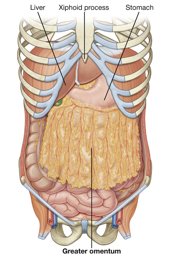
The xiphoid process and costal. Having visible abs is a byproduct of nutrition, exercise, and overall caloric expenditure. Abdominal computed tomography (ct) is a type of medical imaging procedure used to diagnose and monitor internal stomach issues, like cancer, bowel obstruction, and abdominal pain. Abdominal anatomy seen on ct. Every day, millions of gym goers do crunches in hopes of getting a tighter, smaller waist.
Abdomen Basicmedical Key from basicmedicalkey.com A collection of anatomy notes covering the key anatomy concepts that medical students need to learn. Abdominal anatomy seen on ct. Laterally by the midaxillary line; There are multiple anatomical areas within the abdomen, each of which contain specific contents and are bound by certain borders. Compare and contrast the different medical imaging modalities presented in the tutorials. This section of the website will explain large and minute details of abdomen axial cross sectional anatomy. Sectional anatomy the sonographer must have a working knowledge of anatomical structures with particular attention to spatial relationships within the body. Who better to review abdominal anatomy with, than an experienced expert?
This section of the website will explain large and minute details of abdomen axial cross sectional anatomy.
The abdominal divisions should be used in conjunction with other diagnostic approaches in order to accurately diagnose a patient's condition. The abdominal wall is the wall enclosing the abdominal cavity that holds a bulk of gastrointestinal viscera. A collection of anatomy notes covering the key anatomy concepts that medical students need to learn. The anterior abdominal wall (figs. Transversus abdominis muscle internal abdominal oblique muscle rectus abdominis muscle external abdominal oblique muscle pyramidalis muscle. Learn about abdominal organs anatomy with free interactive flashcards. Most students entering ultrasound have some basic understanding of anatomy. Every day, millions of gym goers do crunches in hopes of getting a tighter, smaller waist. Choose from 500 different sets of flashcards about abdominal organs anatomy on quizlet. Free and interactive atlas of the human anatomy. Laterally by the midaxillary line; Radiographers suggest an abdominal ct scan to look for the following 5 name the nine abdominal regions and their main contents.
The xiphoid process and costal. 6 write the origin, insertion and nerve supply of muscles of anterior abdominal wall. The abdomen (colloquially called the belly, tummy, midriff or stomach) is the part of the body between the thorax (chest) and pelvis, in humans and in other vertebrates. The abdomen refers to the region between the pelvis (pelvic brim) and the thorax (thoracic diaphragm) in vertebrates, including humans. The above lines intersect and divide the abdomen into nine regions (clockwise from the top)
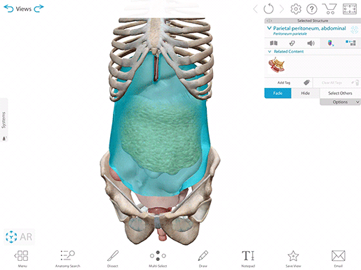
Studying The Peritoneum With Human Anatomy Atlas 2020 from www.visiblebody.com Who better to review abdominal anatomy with, than an experienced expert? In anatomy and physiology, you'll learn how to divide the abdomen into nine different regions and four different quadrants. Understanding abdominal anatomy and physiology is essential to understanding the human body as a whole. And inferiorly by the symphysis pubis, pubic tubercle, inguinal ligament, anterior superior iliac spine, and. The abdomen (colloquially called the belly, tummy, midriff or stomach) is the part of the body between the thorax (chest) and pelvis, in humans and in other vertebrates. Demonstrate comprehension of core abdominal anatomy. Radiology basics of abdominal ct anatomy with annotated coronal images and scrollable axial images to help medical students and junior doctors learning anatomy. The problem is that the basic crunch is.
Demonstrate comprehension of core abdominal anatomy.
Compare and contrast the different medical imaging modalities presented in the tutorials. This muscle forms the anterior and lateral abdominal wall. Radiographers suggest an abdominal ct scan to look for the following Demonstrate comprehension of core abdominal anatomy. Having visible abs is a byproduct of nutrition, exercise, and overall caloric expenditure. • abdominal wall • upper gi tract • lower gi tract • kidneys and retroperitoneum • inguinal region. Abdominal anatomy seen on ct. Lee moffitt cancer center & research institute in. The abdomen (colloquially called the belly, tummy, midriff or stomach) is the part of the body between the thorax (chest) and pelvis, in humans and in other vertebrates. Every day, millions of gym goers do crunches in hopes of getting a tighter, smaller waist. Learn about abdominal organs anatomy with free interactive flashcards. This mri abdomen axial cross sectional anatomy tool is absolutely free to use. Identify some abdominal pathology on medical images.
In anatomy and physiology, you'll learn how to divide the abdomen into nine different regions and four different quadrants. Every day, millions of gym goers do crunches in hopes of getting a tighter, smaller waist. Who better to review abdominal anatomy with, than an experienced expert? • in this module, we will explore basic abdominal anatomy identifiable with common imaging modalities. The anterior abdominal wall (figs.
Understanding Abdominal Divisions Anatomy Snippets Complete Anatomy from s3-us-west-1.amazonaws.com A collection of articles covering abdominal anatomy, including abdominal wall anatomy and abdominal cavity anatomy. The abdominal divisions should be used in conjunction with other diagnostic approaches in order to accurately diagnose a patient's condition. In anatomy and physiology, you'll learn how to divide the abdomen into nine different regions and four different quadrants. Introduction to sonographic abdominal anatomy. Radiology basics of abdominal ct anatomy with annotated coronal images and scrollable axial images to help medical students and junior doctors learning anatomy. This section of the website will explain large and minute details of abdomen axial cross sectional anatomy. Abdominal surface anatomy can be described when viewed from in front of the abdomen in 2 ways: Divided into 9 regions by two vertical and two horizontal imaginary planes.
The abdominal divisions should be used in conjunction with other diagnostic approaches in order to accurately diagnose a patient's condition.
Divided into 9 regions by two vertical and two horizontal imaginary planes. Lee moffitt cancer center & research institute in. In anatomy and physiology, you'll learn how to divide the abdomen into nine different regions and four different quadrants. Introduction to sonographic abdominal anatomy. Having visible abs is a byproduct of nutrition, exercise, and overall caloric expenditure. The xiphoid process and costal. The abdominal divisions should be used in conjunction with other diagnostic approaches in order to accurately diagnose a patient's condition. A collection of articles covering abdominal anatomy, including abdominal wall anatomy and abdominal cavity anatomy. Prep for a quiz or learn for fun! We created an anatomical atlas of abdominal and pelvic ct which is an interactive tool for studying the conventional anatomy of the normal structures based. A good amount of area is covered by the abdominal wall. 6 write the origin, insertion and nerve supply of muscles of anterior abdominal wall. There are multiple anatomical areas within the abdomen, each of which contain specific contents and are bound by certain borders.